Overview
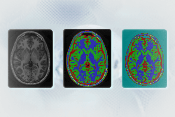
This project focuses on automating the segmentation of 3D medical images, such as MRI scans, using a combination of Otsu’s thresholding and K-means clustering algorithms. The objective was to create a robust, open-source solution capable of handling complex medical images, which often exhibit overlapping intensity distributions. The implementation, developed in C++, leverages the Insight Segmentation and Registration Toolkit (ITK) and Visualization Toolkit (VTK) libraries for image processing, segmentation, and visualization. The system is designed to assist in precise, automated image analysis for medical professionals and researchers.
Core Challenges and Approach
Medical image segmentation is critical for isolating specific anatomical structures, but it poses unique challenges due to image noise, intensity inhomogeneity, and subtle variations in tissue contrast. This project addressed these challenges by integrating two complementary algorithms:
Otsu’s Thresholding Method: A global thresholding technique that automatically computes the optimal threshold to segment images into distinct classes by maximizing the variance between them. It is particularly suited for distinguishing tissues with clear intensity differences.
K-means Clustering: After the initial segmentation with Otsu’s method, K-means clustering was applied iteratively to refine the segmentation boundaries. This unsupervised learning algorithm assigns each voxel to a class based on its intensity and iteratively recalculates the mean intensity for each class, improving the precision of the segmentation. This method is essential for adjusting the segmentation in areas where Otsu’s method falls short, particularly in regions with overlapping intensities.
Implementation Details
ITK Integration: The code is written in C++ and utilizes ITK for medical image processing. The pipeline begins with reading the input medical image, followed by applying Otsu’s multiple threshold filter. The image is segmented into K classes (clusters), with the optimal value of K being determined through experimentation based on the dataset.
K-means Clustering Refinement: After initial segmentation, the algorithm performs K-means clustering to fine-tune class boundaries. This iterative process continues until the change in mean intensities between clusters falls below a predefined threshold or until the maximum number of iterations is reached. The number of clusters K is passed as a command-line argument, allowing for flexibility in defining segmentation granularity.
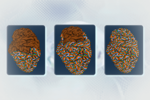
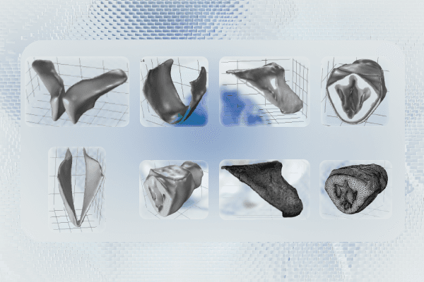
Real-Time Visualization with VTK: VTK is used for rendering and visualizing the segmented data in real-time. The program generates a 3D mesh from the segmented image, and a custom callback function updates the visualization at regular intervals, simulating a diffusion-like process across the mesh. This real-time interaction aids in understanding the segmentation process and assessing the accuracy of the algorithm dynamically.
Technical Features
Dynamic Segmentation: The segmentation process allows for dynamic adjustments of the number of clusters (K) based on the characteristics of the medical image, making it adaptable to various datasets.
Real-Time Rendering: Using VTK’s powerful rendering capabilities, the segmented mesh is visualized with dynamic updates that display changes in vertex colors as the segmentation or diffusion process progresses, providing immediate visual feedback.
Scalability: The system is designed to handle large 3D medical datasets efficiently, thanks to optimized data structures in ITK and VTK for voxel-level operations and mesh traversal.
Performance and Computational Efficiency
Timer-Based Animation: The diffusion process used in the system is controlled by a timer, which triggers the execution of the next iteration of the segmentation process. This not only allows real-time visualization but also ensures that the computational load is distributed over time, maintaining responsiveness during rendering.
Computational Optimization: To improve the overall computational efficiency, a lookup table is employed for rapid color mapping during the visualization process. Furthermore, efficient neighbor-detection algorithms from VTK are leveraged to optimize the traversal of large voxel datasets, reducing the time complexity of the segmentation and diffusion process.
Alternative Approaches: The project explored several alternative techniques, including event-driven and multi-threaded processing approaches, to enhance performance for more complex or larger medical datasets. Advanced neighbor detection algorithms like kd-trees and spatial hashing were also considered to optimize the search space for neighboring voxels, further improving computational efficiency.
Key Technical Contributions
Hybrid Segmentation Framework: By combining Otsu’s method for initial segmentation and K-means clustering for refinement, the system offers a balanced approach that integrates global thresholding with localized clustering to produce high-accuracy segmentations.
Configurable Granularity: The segmentation granularity, controlled by the value of K (number of clusters), allows users to fine-tune the segmentation according to the specific clinical or research requirements, whether they need coarse or highly detailed segmentations.
Real-Time 3D Visualization: The system’s ability to visualize 3D mesh data in real time as it evolves through each iteration of the diffusion process offers valuable insights into the segmentation’s effectiveness and helps identify potential areas for improvement.
Outcome
This project successfully developed an automated segmentation tool that can accurately and efficiently process complex 3D medical images. The combination of Otsu’s thresholding and K-means clustering provided a powerful hybrid method for medical image segmentation, offering flexibility and adaptability for different medical imaging scenarios. The tool’s real-time visualization capabilities make it easier to evaluate segmentation accuracy and adjust parameters as needed, providing clinicians and researchers with a valuable tool for medical image analysis.
